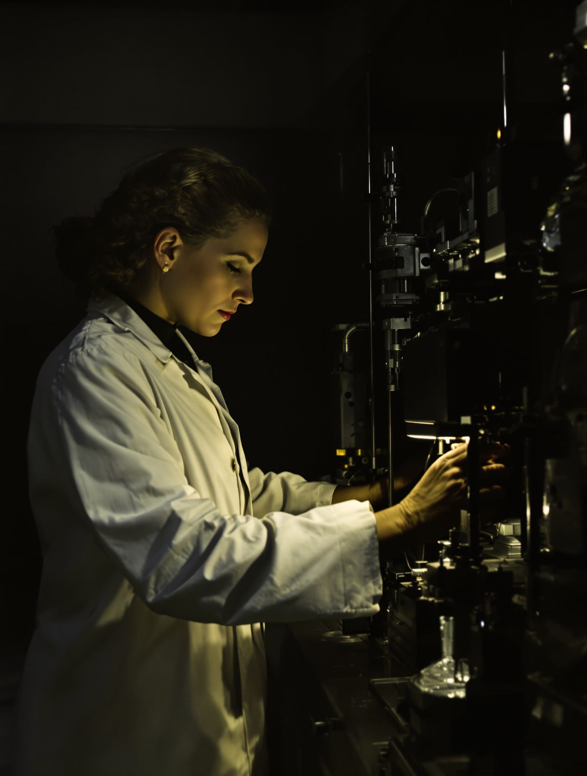
In the basement of King's College London, surrounded by the hum of machinery and the acrid smell of photographic chemicals, Rosalind Franklin peered through the viewfinder of her X-ray camera. It was May 1951, and she had no idea she was about to capture the most important photograph in the history of science—an image that would unlock the secret code of life itself.
The strand of DNA suspended in her apparatus was thinner than a human hair, invisible to the naked eye. Yet through her revolutionary technique, Franklin was about to reveal its hidden architecture with startling clarity. What emerged from her darkroom that day would be dubbed "Photo 51"—a grainy, black-and-white image that looks unremarkable to untrained eyes but contained within its shadows and bright spots the blueprint for understanding heredity, evolution, and the very essence of what makes us human.
The Woman Who Saw in X-Rays
Rosalind Elsie Franklin arrived at King's College London in January 1951, aged just 30, but already armed with a formidable reputation. She had spent four years in Paris perfecting the art of X-ray crystallography—a technique that fires X-ray beams through crystalline substances to reveal their atomic structure. While her male colleagues at King's were still fumbling with crude DNA samples, Franklin brought Parisian precision to the problem.
Her laboratory in the basement was a shrine to meticulous preparation. Where others saw tedious detail, Franklin saw the difference between breakthrough and failure. She didn't just point X-rays at DNA and hope for the best—she controlled humidity with obsessive care, knowing that DNA changes shape as it absorbs water from the air. This attention to detail would prove crucial.
Franklin had identified two distinct forms of DNA: the dry "A-form" and the wet "B-form." Most researchers worked with whatever DNA they happened to have, but Franklin realized that the B-form—DNA in its natural, hydrated state—held the key to understanding how it actually functioned in living cells.
100 Hours to Capture Life's Blueprint
The creation of Photo 51 was an exercise in extraordinary patience. Franklin positioned her DNA sample—a single fiber extracted from calf thymus—in her custom-built X-ray apparatus. The process required absolute stillness: the tiniest vibration would blur the image and waste hours of work.
For more than four days, her machine bombarded the DNA with X-ray beams while a photographic plate captured the scattered rays. Each hour of exposure revealed a little more detail, like a Polaroid slowly developing in reverse. Franklin couldn't peek at her progress—opening the camera would ruin everything.
The technique itself bordered on alchemy. X-rays, passing through the DNA's molecular structure, scattered in patterns that corresponded to the arrangement of atoms within. Dense areas of the molecule blocked more rays, creating dark spots on the photographic plate. Sparse areas let rays through, creating bright regions. The resulting image was essentially a shadow-map of DNA's architecture.
When Franklin finally developed Photo 51 in May 1951, she must have caught her breath. The image showed something unprecedented: a clear X-pattern in the center, indicating a helical structure, surrounded by diamond-shaped patterns that revealed the spacing between molecular layers. Even more remarkably, the symmetry of the image suggested that DNA consisted of not one, but two intertwined helical chains.
The Photograph That Almost Stayed Secret
What happened next would become one of science's most controversial episodes. Franklin, cautious by nature, didn't immediately publish her findings. She wanted more data, more certainty. Photo 51 was extraordinary, but she was a perfectionist working in a field where premature conclusions could destroy careers.
Her colleague Maurice Wilkins, however, had other ideas. Without Franklin's knowledge or consent, Wilkins showed Photo 51 to James Watson, a young American biologist working with Francis Crick at Cambridge University. The pair had been struggling to understand DNA's structure using theoretical models—but Franklin's photograph gave them the missing pieces of the puzzle.
Watson later described the moment he first saw Photo 51 as electrifying: "The instant I saw the picture my mouth fell open and my pulse began to race." The X-pattern screamed "helix" to anyone who understood crystallography. The precise measurements Franklin had calculated told them exactly how wide the helix was and how far apart its molecular rungs sat.
Within weeks, Watson and Crick had built their famous double helix model—using Franklin's data as their foundation. When they published their breakthrough in Nature in April 1953, Franklin's crucial contribution was buried in a footnote, her photograph referenced only obliquely.
The Hidden Tragedy Behind the Triumph
The true tragedy of Rosalind Franklin's story isn't just that she was denied proper credit—it's that she was so close to solving the puzzle herself. Her laboratory notebooks, examined decades later, show she had identified the double helix structure independently. In her notes from early 1953, she wrote: "Structure B is probably a helical structure with 2, 3 or 4 co-axial nucleic acid chains."
She had also deduced that DNA's phosphate groups faced outward (contradicting Watson and Crick's initial model) and that the two chains ran in opposite directions. These insights were crucial to understanding how DNA actually works—yet they emerged from painstaking analysis of her X-ray images rather than theoretical speculation.
Franklin's meticulousness, often criticized as excessive caution, was actually producing more accurate results than the Cambridge team's bold model-building. But in the race for scientific immortality, careful analysis lost to confident publicity.
Tragically, Franklin died of ovarian cancer in 1958, aged just 37—possibly caused by her years of exposure to X-rays at a time when safety protocols were primitive. When Watson, Crick, and Wilkins received the Nobel Prize in 1962 for discovering DNA's structure, Franklin was no longer alive to share the honor. Nobel Prizes aren't awarded posthumously, but many historians argue she would have been excluded anyway, such was the male-dominated culture of 1960s science.
The Photograph That Changed Everything
Photo 51 didn't just reveal DNA's structure—it opened the door to the molecular age. Understanding the double helix led directly to breakthroughs in genetics, the discovery of how genes encode proteins, and eventually to technologies we now take for granted: DNA fingerprinting, genetic engineering, and the Human Genome Project.
Every time scientists identify a disease gene, develop a new vaccine, or trace human migration patterns through genetic analysis, they're building on the foundation that Franklin's photograph provided. The COVID-19 vaccines that emerged with unprecedented speed? They exist because we understand how genetic code works—knowledge that traces back to that basement laboratory at King's College.
Today, Photo 51 hangs in the Science Museum in London, a grainy testament to one of history's greatest scientific achievements. Visitors often walk past without realizing they're looking at the image that unlocked the secret of heredity—the photograph that revealed how parents pass traits to children, how evolution shapes species, and how life itself copies and perpetuates itself across generations.
Rosalind Franklin's legacy reminds us that scientific breakthroughs rarely emerge from sudden inspiration alone. They grow from meticulous work, patient observation, and the courage to peer into the unknown armed with nothing but curiosity and precision instruments. In capturing Photo 51, Franklin didn't just photograph DNA—she captured the moment humanity first glimpsed the molecular foundation of life itself. That grainy image from a London basement remains one of the most profound human achievements: the day we finally saw ourselves at the atomic level, and understood how wonderfully, precisely, we are made.